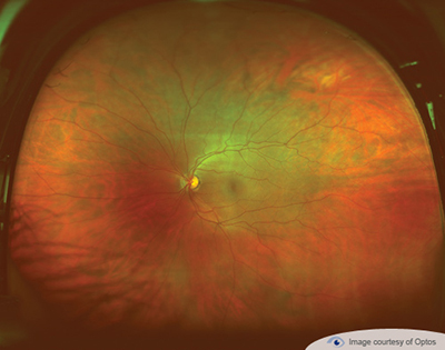

Current research is also showing that ocular melanoma is very different from normal skin melanoma. Other sites include ciliary body, iris, and conjunctiva. FAF is becoming an increasingly useful tool in the diagnosis of numerous pathologies including geographic atrophy, retinitis pigmentosa, stargardt disease, best disease, hydrochloroquine retinopathy and others. Choroidal melanomas are the most common site comprising 85% of cases. Fundus autofluorescence utilizes the natural fluorescent properties of lipofuscin within the retinal pigment epithelium to create an image of the retina. There are other types of eye cancers, but melanoma is the most common. Ocular melanoma is often lethal, but thankfully, a very rare disease. Increased pressure can result in changes to blood vessels in the eye, increasing the risk of cardiovascular disease (stroke or heart disease). Glaucoma causes damage to the optic nerve and almost always develops without symptoms. The results in alterations to our fine central vision making daily activities difficult. The center of the retina (the macula) can become diseased when we get older. Retinopathy occurs when diabetes damages the tiny blood vessels inside the retina. Early detection is essential so treatments can be administered.ĭiabetes affects the eyes and the kidneys and is a leading cause of blindness. This means, in addition to eye conditions, signs of other diseases (such as stroke, heart disease, hypertension and diabetes) can also be seen in the retina. Your retina is the only place in the body where blood vessels can be seen directly. Routine exams were uncomfortable, especially for a child, which made it impossible for the doctor to conduct a complete exam and view the entire retina. In this type, fluid leaks into the area underneath the retina (subretina).Įarly detection of all of these diseases and conditions mean successful treatments can be administered which reduces the risk to your sight and health. Optos was founded over 20 years ago by Douglas Anderson after his then five-year-old son, Leif, went blind in one eye when a retinal detachment was detected too late.

This type of detachment is less common.Įxudative – Frequently caused by retinal diseases, including inflammatory disorders and injury/trauma to the eye. Tractional – In this type of detachment, scar tissue on the retina’s surface contracts and causes it to separate from the RPE. These types of retinal detachments are the most common. Rhegmatogenous – A tear or break in the retina causes it to separate from the retinal pigment epithelium (RPE), the pigmented cell layer that nourishes the retina, and fill with fluid.

What are the different types of retinal detachments? Anyone can get a retinal detachment however, they are far more common in nearsighted people, those over 50, those who have had significant eye injuries, and those with a family history of retinal detachments. Vitreous degeneration (liquefaction or shrinkage), Myopia, Fellow eye with retinal detachment, Strong family history of retinal. If not promptly treated, a retinal detachment can cause permanent vision loss.
#RETINAL DETACHMENT OPTOMAP PROFESSIONAL#
Some patients, however, will need more than one procedure to repair the damage.Īn eye care professional who has examined the patient's eyes and is familiar with his or her medical history is the best person to answer specific questions.When the retina detaches, it is lifted or pulled from its normal position. Please note that optomap Retinal Imaging services are not available at all eyecarecenter locations. Salt solution is then injected to into the eye to replace the vitreous.Įarly treatment can usually improve the vision of most patients with retinal detachments. detachments, and other health problems such as diabetes. Next, a small instrument is placed into the eye to remove the vitreous. During a vitrectomy, the doctor makes a tiny incision in the sclera (white of the eye). If necessary, a vitrectomy may also be performed to treat more severe cases. In some cases a scleral buckle, a tiny synthetic band, is attached to the outside of the eyeball to gently push the wall of the eye against the detached retina.

Retinal detachments are treated with surgery that may require the patient to stay in the hospital. If you cannot find an optomap location in your area, please contact us. Cryopexy is a similar procedure that freezes the area around the hole. Retinal Detachment optomap Story Find optomap locations: Results. Retinal detachment: The main cause of this. During laser surgery, tiny burns are made around the hole to "weld" the retina back to into place. Retinal tear: This occurs as a result of pulling the transparent and jelly-like substance (vitreous) on the retina. These procedures are usually performed in the doctor's office. Small holes and tears are treated with laser surgery or a freeze treatment called cryopexy.


 0 kommentar(er)
